HOW ARE AIRWAY & POSTURE RELATED?
When we think of "airway, we think of life-and-death situations and sleep apnea. But, there is so much more to our ability to pull air into our lungs freely and evenly and to expel carbon dioxide. Our alignment and muscle tone play a pivotal role in our ability to maintain a symmetrical, un-restricted airflow. You see, the respiratory system is a body-wide, interconnected and closed system. There are often clues our body is giving us that there is a restriction somewhere to free, full airflow. Often times, the restrictions are located quite a distance from our nose and mouth!
Key Points

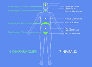
First of all, it is not common knowledge, but we actually have 4 diaphragms (some even say 5) that all influence each other. They act as one closed, pressurized system. If there is an issue with the function of one of the diaphragms, it will influence the others.

Another key concept is that our main diaphragm, the respiratory diaphragm, is actually made up of 2 separate domes - a right and a left dome. They do share a common tendon (called the central tendon), but they are much different in shape and attachment to the spine. This means that just because you are breathing, there is no guarantee that you are using both sides evenly. Not only is it possible to be breathing asymmetrically, it is highly likely!
Now onto some specific, overlooked posture and physical clues that someone may be dealing with an airway problem.
1) Asymmetrical Face:

Believe it or not, the cranial bones actually move. One specific way they move is during every breath. They flex and open when we inhale... and extend and draw in when we exhale (See image to Left). So, if we are struggling to adequately fill one side of our body with air, the skull will show it. In the same way, if we are overfilling one side of our body with air, the skull will show it.

 What could it look like?

More Fullness on One Side (Zygomatic Region)
More Indentation of Temporal Region on One Side
Wider Opening of One Eye
One Nostril Wider and More Open
More Visible Flare of One Ear
One Nostril Wider and More Open
One Corner of the Eye Closer to Side of Face
Uneven Eyebrow Levels
2) Neck Tension:
This one is probably more well known. When we are struggling to open out airway, our neck muscles will jump in to help. Ever noticed runners who hold their shoulders up toward their ears? Or a really muscular neck? This is a desperate attempt by the neck muscles to make up for a lack of full airway opening. They will elevate the first few ribs to allow more air in.
3) Back Tension:
The diaphragm attaches to our lumbar spine (low back). If there is insufficient movement of the diaphragm, which happens as a result of inappropriate airflow, it will create a compression of our lumbar spine.
In addition, when we are unable to fully expand the back of our lungs (one of the more common restrictions, we will fill at the easiest area... the lower, front lobes. We then over-fill the lower front lobes and allow our abdomen to protrude forward. This will further cause a compression of the low back. Just like the neck will contract to help us get in more air, so do our back muscles. We tense them in order to pull our ribs together in the back, allowing more opening in the front ribs.
4) Uneven Shoulders:

 Since the lungs actually provide 3-D opening and structural support of our ribcage, we often present with one side of the ribcage more closed down and "collapsed" while the other is open and full.
In an Upright Position, this may look like this:

Or This.... As a compensation

Remember, the respiratory diaphragm directly influences the other 3 diaphragms via pressure and pistoning. An imbalance here will create an imbalance in the others. In the same way, we can influence the other diaphragms by balancing this one!
5) Pelvic Floor Issues:

Because the 4 diaphragms work in synchrony, a problem with the pelvic diaphragm's ability to ascend or descend can affect the ability of our other diaphragms to ascend or descend... an vice versa. The pelvic diaphragm should be continually active during all phases of respiration. During Inhalation, the Pelvic Diaphragm descends. During Exhalation, it ascends. A chronically descended pelvic diaphragm corelates with an increased amount of Respiratory Inhalation (or hyper-inflation). Likewise, if the pelvic diaphragm is limited in it's ability to descend (or it's stuck in an ascended state), it will LIMIT the ability to inhale fully with respiration. Pelvic floor relaxation = inhale. Pelvic flor contraction = exhale
What is common to see is a pelvic diaphragm that is asymmetrical. Look closely at the pelvis x-ray here. Notice the different shape of the Obturator Foramen (bottom holes) and the orientation of the ischial rings (the sit bones). The muscles and ligaments that attach to those bones will be pulled, twisted or compressed along with them. Just another example that our airway and pressures can be asymmetrical.
6) Hip Tightness:
There are 2 generalized areas of the hip that can feel tight. The font of the hips (Hip Flexors - Psoas, Iliacus, Anterior Pelvic Floor); the sides and back of the hips (Obturator Internus, Posterior Pelvic Floor, Piriformis, TFL/Glute Medius/Glute Minimus).
Let's take a look at the front of the hips first. One key point here is the fact that the Psoas muscle attaches to our lumbar spine and is directly connected to our diaphragm via the thick tendon-like fascia often called the Psoas Fascia. The other end of the Psoas is at the upper femur (thigh bone). This connection of the diaphragm to the front of the femur explains why tight hip flexors can reflect a breathing dyssynchrony.

To understand how respiration and airflow are related to lateral and posterior hip tightness, take a look at how the Piriformis and Obturator Internus form the walls of the pelvic openings, aiding in resisting pressure. The Obturator Internus even attaches to the pelvic diaphragm directly, via a strong fascia band.

Take a look at that thickness! Remember the pelvic diaphragm's movement relative to the thoracic
diaphragm? The Obturator Internus will lengthen during inhalation and shorten during exhalation. Therefore, a restriction in airflow/pressure imbalance at the pelvic diaphragm will influence tone and position of the Obturator Internus (among others)… and vice-versa. Likewise, overall pressures in the pelvic floor will affect the position and tone of the Piriformis.
To even further understand this relationship, we must follow these muscles to their lateral insertion. Notice where these 2 muscles attach to the outer femur, and very close to each other. In a nutshell, the pelvic diaphragm and outer hip are highly linked!
Relationship of Our 4 Diaphragms


Comments
Post a Comment